Who We Are
At the Coleen Cunningham Foundation (CCF), we offer heartfelt support and vital resources to individuals and families facing atypical Parkinsonism. Our mission is to uplift lives through compassionate care—providing respite and hospice grants, support groups, and advocacy. We’re dedicated to building a community that inspires hope, fosters understanding, and strengthens resilience, empowering patients and caregivers to face this journey with dignity and connection.
Useful Information
-

On the Journey - Weekly Zoom Support Group
LEARN MOREThis Meeting is open to All; Caregivers, Individuals, Healthcare Workers and Loved Ones.
Every Thursday 7-9 pm ET
Attendees from all across the globe join us for support and to raise awareness.
-

Faces & Voices of Atypical Parkinsonism
LEARN MOREShare your personal story with Atypical Parkinsonism, including PSP, MSA, CBD, and DLB, whether you are diagnosed, a caregiver, family member, friend, or medical professional. Together, we can light the path toward understanding and hope.
-

Events & Fundraising
VIEW EVENTSCCF hosts and participates in awareness walks and fundraising initiatives to raise funds for Atypical Parkinsonism research and support services, benefiting those with PSP, MSA, CBD, and DLB.
Over $45,000 Raised!
-

Discover Vielight Red Light Therapy for Atypical Parkinsonism
MORE INFOLiving with Atypical Parkinsonism presents unique challenges, but innovative solutions like Vielight red light therapy can help. This cutting-edge, non-invasive treatment uses red and near-infrared light to support brain health, enhance mood, and improve quality of life for those with Atypical Parkinsonism.


Support CCF
Your Donations to CCF support our Programs for Individuals and Families touched by Atypical Parkinsonism. With your donations we are able to offer support and resources to those in need, and make sure No One Walks Alone on this journey.
Fundraisers
-
 START TODAY
Support CCF: Unleash Your Fundraising Power with Donorbox
Create a Fundraiser in a Few Steps.
Whether it’s for a special occasion, a personal challenge, or in memory of a loved one, you can set up your own fundraising page in minutes. Once it’s live, simply share the link with your friends, family, and network to start raising funds for CCF. -
 MORE INFO
2026 Global Atypical Parkinsonism Awareness Walk
Get ready to lace up your shoes and join a worldwide movement! CCF is thrilled to announce the 2026 Global Awareness Walk, expanding on our successful annual events to unite communities across the globe in support of PSP and Atypical Parkinsonism (MSA, CBD, and DLB).


Understanding Atypical Parkinsonism
Find the information, support, and inspiration you need to better understand and manage these challenging conditions. Our library includes medical guides, personal memoirs, and practical advice to help patients, caregivers, and families navigate the complexities of PSP, MSA, CBD.
Resources
-

Support & Resource Links - Worldwide
WORLDWIDE LINKS -
New Symptom Treatment - VieLight - Red Light Therapy
LEARN MORE -

Supports & Resource Links - Canada
CANADIAN LINKS -

Assistive Device Exchange
VIEW LISTINGS -

Tim Talks PSP/CBD Warrior
VIEW RECORDINGS -

Awareness Month Downloads
GET YOURS HERE


Atypical Parkinsonism Blog by: Laura Louizos
View all-

Remembering Rev. Jesse Jackson: A Warrior’s Fig...
We at CCF for PSP Awareness are deeply saddened by the passing of Rev. Jesse Jackson, civil rights pioneer, at age 84 on February 17, 2026, in Chicago. His death...
Remembering Rev. Jesse Jackson: A Warrior’s Fig...
We at CCF for PSP Awareness are deeply saddened by the passing of Rev. Jesse Jackson, civil rights pioneer, at age 84 on February 17, 2026, in Chicago. His death...
-
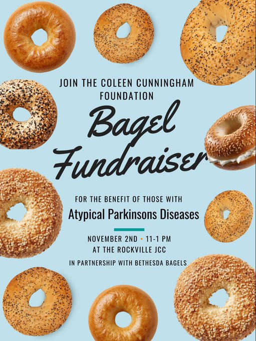
🥯 Bagel Fundraiser
JOIN THE COLEEN CUNNINGHAM FOUNDATION 🥯 Bagel Fundraiser FOR THE BENEFIT OF THOSE WITHAtypical Parkinson Diseases NOVEMBER 2ND • 11-1 PM AT THE ROCKVILLE JCC IN PARTNERSHIP WITH BETHESDA BAGELS
🥯 Bagel Fundraiser
JOIN THE COLEEN CUNNINGHAM FOUNDATION 🥯 Bagel Fundraiser FOR THE BENEFIT OF THOSE WITHAtypical Parkinson Diseases NOVEMBER 2ND • 11-1 PM AT THE ROCKVILLE JCC IN PARTNERSHIP WITH BETHESDA BAGELS
-

HOPE by Bhavna
Remember the first timerobins sang in your garden,their delicate bodies pulsing with song,their hearts daring to rise, like yours.How the moon spilled its silver over your window,and your mother said,...
HOPE by Bhavna
Remember the first timerobins sang in your garden,their delicate bodies pulsing with song,their hearts daring to rise, like yours.How the moon spilled its silver over your window,and your mother said,...


Support CCF
Your Donations to CCF support our Programs for Individuals and Families touched by Atypical Parkinsonism. With your donations we are able to offer support and resources to those in need, and make sure No One Walks Alone on this journey.